Many of us have heard of MRI, which stands for magnetic resonance imaging – a non-invasive technique that uses a scanner to produce vivid images of the human brain. But most researchers aren’t interested in static images; they are trying to understand dynamic, real-time activity in various regions of the brain. That’s where functional MRI, or fMRI, comes into play. While a subject is lying in the scanner, they can see a computer screen that will play any number of stimuli. Depending on the Marketer’s and Researcher’s hypotheses, subjects can be shown a video (such as a commercial), a series of images (of various products or logos), or a cognitive task (like a decision making paradigm). Subjects are also fitted with headphones to hear any instructions or auditory stimuli that accompany the visual images, and also have access to a button press apparatus to record their responses to audiovisual tasks.
An fMRI obtains images by sending radio waves within the core of an incredibly strong electromagnet – which is on average 50,000 times greater than the Earth’s magnetic force. Once a person enters the magnetic field, the proton molecules in their bodies line up. Protons are electrically charged particles that are found in the nuclei of oxygen atoms, which play an important part in producing the images captured during fMRI. Just like any other organ or muscle in the body, when a given brain region is put to use it receives an increased supply of oxygen rich blood to “feed” the millions of neurons hard at work. The fMRI sends a radio wave pulse to knock the protons out of their configuration, and when they line up again the protons release a signal that is read by the scanner. The resulting blood-oxygen-level-dependent, or BOLD signal is what allows Neuromarketers and Researchers to know which brain regions are responsible for processing an image of a preferred shirt colour, or emotional responses to a song heard during a commercial. Radio pulses can be sent every one or two seconds, which provide an output of multiple images that can be analyzed for changes in BOLD signal over time.
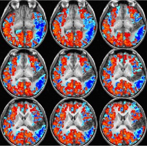
Sample of visualized fMRI imaging data:
Some of the benefits of fMRI include the great resolution that is provided from resulting images, and that unlike scalp recordings from EEG, fMRI can obtain information from areas deep within the human brain which process emotion and automatic reflex responses. The magnetic field produced by the scanner is safe, except for those with any non-removable metal in their bodies including metal fillings, pins, and pacemakers. However, many metals like aluminum are non-ferromagnetic, which means they won’t be attracted to a magnetic force, although they will create artifacts or ‘bright spots’ in the resulting images. This is usually seen with subjects with non-removable retainers, who can be scanned with fMRI but will usually ‘outshine’ any activity in brain regions close to their mouth. It is also not recommended to scan pregnant women, whose brains can be scanned with other techniques like EEG should the need arise.
Safety certified technicians are present, and all consumer neuroscience experiments are been approved by an ethics committee before testing can begin, which speeds up the timeline of the Neuromarketing research study. The insights emerging from fMRI are proving to far outweigh the technical considerations of this neuroimaging technique. In future blog posts, we’ll discuss the specific benefits and measures derived from fMRI consumer research.